Introduction
Diabetes mellitus disrupts the body’s natural wound‑healing process, often leading to persistent, non‑healing wounds especially diabetic foot ulcers (DFUs) that severely impact quality of life and healthcare costs. With roughly 19–34% of diabetics developing DFUs during their lifetime [14], it's vital to understand the biology of delayed healing in diabetes and explore effective management strategies. This article uncovers key pathophysiological mechanisms and emerging treatments rooted in basic science and clinical research.
Phases of Normal Wound Healing vs. Diabetic Impairment
Wound healing is a highly coordinated biological process that involves four overlapping phases: hemostasis, inflammation, proliferation, and remodeling. Each phase is critical, and any disruption—like that caused by diabetes—can delay or completely derail healing. Let’s break down each phase and explore how it’s impaired in diabetes:
1. HEMOSTASIS PHASE
What normally happens:
Right after injury, blood vessels constrict and platelets aggregate to form a fibrin clot. This stops bleeding and forms the temporary matrix for cell migration.
What happens in diabetes:
Platelet function is often impaired, and clot formation may be delayed or unstable [1]. In addition, hyperglycemia can alter the fibrin structure, making the clot more resistant to degradation and delaying transition to the next phase [2].
2. INFLAMMATORY PHASE
What normally happens:
Within hours, immune cells like neutrophils and macrophages arrive at the wound site to clear pathogens, debris, and damaged tissue. This phase sets the stage for repair.
What happens in diabetes:
In diabetic wounds, inflammation is prolonged and dysregulated. Neutrophils produce excessive reactive oxygen species (ROS) and form neutrophil extracellular traps (NETs), which damage surrounding tissues [3]. Macrophages fail to switch from the inflammatory M1 to the healing M2 phenotype, sustaining a pro-inflammatory environment [4].
3. PROLIFERATION PHASE
What normally happens:
New tissue starts to form. Fibroblasts deposit collagen, endothelial cells promote angiogenesis, and keratinocytes migrate to cover the wound.
What happens in diabetes:
This phase is significantly compromised. Angiogenesis is impaired due to reduced VEGF expression and endothelial dysfunction [5]. Fibroblasts exhibit reduced proliferation and altered collagen production [6]. Keratinocyte migration and re-epithelialization are delayed, often due to disrupted signaling and cytoskeletal abnormalities [7].
4. REMODELING (MATURATION) PHASE
What normally happens:
Over weeks to months, the wound matures. Collagen is reorganized (type III replaced by type I), unnecessary vessels are removed, and tensile strength improves.
What happens in diabetes:
Collagen remodeling is inefficient, there’s excessive type III collagen and insufficient cross-linking [8]. As a result, the scar remains weak and the wound site is prone to re-injury. Matrix metalloproteinases (MMPs) remain overexpressed, degrading new tissue instead of stabilizing it [9].
Key Pathophysiological Mechanisms
a) Chronic Hyperglycemia & Oxidative Stress
Persistent high glucose levels promote the formation of advanced glycation end-products (AGEs), reduce antioxidant enzymes like glutathione peroxidase and superoxide dismutase, and elevate reactive oxygen species (ROS) that damage cells, microvascular endothelium, and nerves, hindering healing [3,17,18].
High ROS also prolongs inflammation and impairs later repair stages [3,19].
b) Impaired Angiogenesis & Endothelial Dysfunction
Healing wounds require new blood vessel growth (angiogenesis) mediated by VEGF and other growth factors. In diabetes, endothelial cells are dysfunctional, VEGF signaling is compromised, and capillary formation is insufficient, leading to chronic ischemia and delayed healing [5,10,25].
c) Neuropathy & Microvascular Compromise
Peripheral neuropathy (sensory, motor, autonomic) leads to reduced nerve density, poor microcirculation, skin dryness, pressure injury, and diminished sensation, increasing DFU risk and delaying detection and healing [3,24].
d) Chronic Inflammation & Immune Dysfunction
Diabetes alters immune cell behavior: neutrophils form excessive NETs, macrophages remain in a pro-inflammatory M1 state, and cytokine balance is disrupted, maintaining a damaging environment and inhibiting progression to repair [13].
e) Extra Cellular matrix & Keratinocytes dysfunction
Hyperglycemia disrupts ECM components and fibroblast activity, impairing collagen deposition. Keratinocyte migration and differentiation are also affected, as seen by reduced laminin-5 and keratin proteins, hindering re-epithelialization [2].
f) Metabolic Aberrations: Insulin Resistance, Lipid & Amino Acid Imbalance
Abnormal metabolism in diabetes affects energy supply, growth factor signaling, and cell function. Insulin resistance impairs fibroblast proliferation and IGF signaling; lipid peroxidation leads to ferroptosis; and arginine deficiency limits nitric oxide (NO), vital for angiogenesis and vasodilation [6,15].
3. Clinical Impact: Diabetic Foot Ulcers
DFUs illustrate the culmination of these pathologies. They develop in 19–34% of diabetic individuals and have high rates of complications and mortality. 60% heal in ~6 months, but 5–24% lead to amputation within 6–18 months [14]. The triad of hyperglycemia, neuropathy, and vascular insufficiency lies at the heart of DFU pathogenesis [14].
4. Management Strategies
A. CONVENTIONAL APPROACHES
Glycemic Control: Maintaining optimal blood glucose levels is foundational, it slows AGE accumulation, oxidative stress, and immune impairment [12,15].
Off‑loading & Pressure Relief: Total Contact Casting (TCC) redistributes pressure away from the ulcer, recognized as the standard for DFUs [23].
Debridement & Infection Control: Regular removal of necrotic tissue and targeted antimicrobial therapy reduce bacterial load and support granulation [1,14].
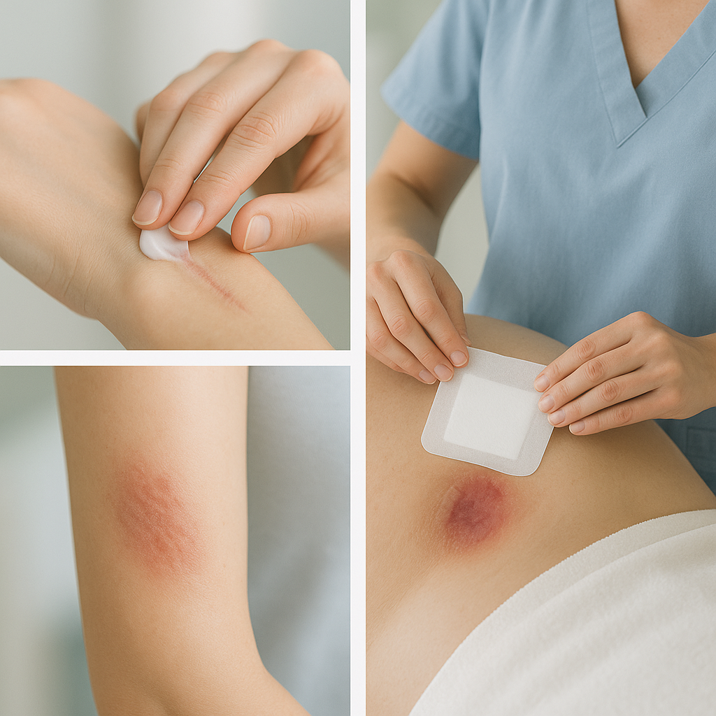
Moist Wound Healing & Dressings: Hydrogel dressings provide moisture balance, oxygen diffusion, and infection resistance suitable for chronic wounds [20].
Negative‑Pressure Wound Therapy (NPWT): Vacuum-assisted closure enhances blood flow, reduces edema, modulates inflammation, and stimulates growth factors, though evidence is mixed [12,21,24].
Revascularization: Correcting macrovascular ischemia with angioplasty or bypass improves perfusion and healing potential [1].
Hyperbaric Oxygen Therapy (HBOT): HBOT may reduce short-term risk of amputation, though benefits past six weeks are unconfirmed [24].
B. ADVANCED & EMERGING THERAPIES
Topical Growth Factors & Bioengineered Skin: VEGF, PDGF, and fibroblast growth factors enhance angiogenesis and repair; extracellular matrix proteins and skin substitutes support cellular growth [1,2,14].
Stem Cells & Gene Therapy: Mesenchymal stem cells and gene delivery of VEGF or neuropeptides show promise in preclinical models [2].
Repurposed Drugs: DPP‑4 inhibitors, metformin, statins, and phenytoin show potential to accelerate healing, though more trials are required.
Hydrogels & Nanotechnology: Advanced hydrogels deliver drugs or respond to redox status; nanodiamond-silk membranes offer temperature sensing and antimicrobial activity [22,28].
Nitric Oxide Dressings & Iron Chelators: NO‑generating dressings and deferoxamine promote vasodilation and angiogenesis by modulating iron and oxidative pathways [14].
Macrophage Modulation: Creams that shift macrophage populations from M1 to M2 improve inflammation resolution and microenvironment [14].
Maggot Debridement & Biologics: Medical-grade larvae effectively remove dead tissue and reduce bacterial burden [14].
Nutrition & Herbal Supplements: Sufficient protein, vitamins, and plant extracts with antioxidants show supportive benefits in healing [7].
Personalized Medicine & Monitoring Technologies: Biomarker‑driven therapies and non-invasive tools like ultrasound/deep learning are emerging for customized treatment and progress tracking [16,29].
Summary & Future Directions
Diabetes impairs healing through hyperglycemia, oxidative stress, vascular and nerve damage, chronic inflammation, ECM dysfunction, and metabolic imbalance. Standard treatments are glycemic control, debridement, dressings, off‑loading, and remain vital, but advanced modalities (growth factors, NPWT, stem cells, hydrogels, personalized interventions) show strong promise.
Future research must focus on:
Robust clinical trials (advanced dressings, repurposed drugs, biomarker-led therapies)
Personalized care using genetic/proteomic profiling
Scaling up new diagnostics (e.g., thermal sensing, AI-powered imaging)
A combined, science‑driven, patient‑centered approach offers the best hope to turn chronic diabetic wounds into healing success stories.
References
Spampinato SF et al. Treatment of impaired wound healing in diabetes: repurposed drugs. Pharmaceuticals. 2020.
Davis FM et al. Dysfunctional wound healing in DFUs. Curr Diab Rep. 2018;18(1):2.
Delavary BM et al. Pathogenesis and treatment of impaired wound healing in diabetes. Int J Low Extrem Wounds. 2014;13(3).
Blakytny R, Jude EB. Altered molecular mechanisms of diabetic foot ulcers. Int J Low Extrem Wounds. 2009;8(2):95‑104.
Johnson KE, Wilgus TA. VEGF and angiogenesis in cutaneous wound repair. Adv Wound Care. 2014.
Zhang et al. Metabolic aberrations in diabetic wound healing. Cell Death Discov. 2025.
Galgotias Univ review: Diabetic wound healing factors & natural products. 2023.
Kang Huang et al. Angiogenesis during diabetic wound repair. Burns & Trauma. 2025.
Lim YC et al. Proinsulin C-peptide and angiogenesis. J Invest Dermatol. 2015;135(1):269‑78.
Khamaisi M et al. NPWT in diabetic foot ulcers. Diabetes Metab Res Rev. 2017;33(7).
Johnson et al. Diabetic microvascular dysfunction & wounds. PMC9319250. 2022.
Swoboda L, Held J. Impaired wound healing in diabetes. J Wound Care. 2022;31(10):882‑5.
Stachura A et al. Wound healing impairment in db/db mouse model. Int J Mol Sci. 2022;23(15):8621.
Spandidos Publ. Developments in management of DFUs. 2025.
Bentham Science. Diabetic wound healing mechanisms & treatments. 2025.
Eurekaselect. Understanding diabetic wounds: mechanisms & multimodal therapy. 2024.
PMC8539411. Diabetic keratinocytes & oxidative damage. 2021.
Delavary BM et al. The problem of wound healing in diabetes. PMC9319250.
PMC8539411. Role of ROS in wound repair.
Wong SL et al. NETosis in diabetic wounds. JMDH bibliometrics. 2024.
Dovepress JMDH. NPWT clinical trial. 2023.
ArXiv Yang J. Hydrogels in diabetic wound treatment. 2024.
Wikipedia Total Contact Casting.
Wikipedia Diabetic Foot Ulcer.
Wikipedia Vascularisation & VEGF.
ArXiv Gao Z. Redox‑responsive hydrogel. 2021.
PMC7243111. Impaired healing & DPP‑4/metformin/statins. 2020.
ArXiv Khalid A. Nanodiamond‑silk membranes for wound healing. 2020.
ArXiv Schlereth M. AI/ultrasound wound monitoring. 2022.